








Coronary & interventional
Your Angiogram Shows a Blockage: Is It Actually Limiting Blood Flow?
An angiogram can show a coronary narrowing without telling the whole story. Dr. Mehdi Shahriyari explains FFR, iFR and IVUS—and how they inform decisions about medication, stents and bypass surgery.
Direct answer
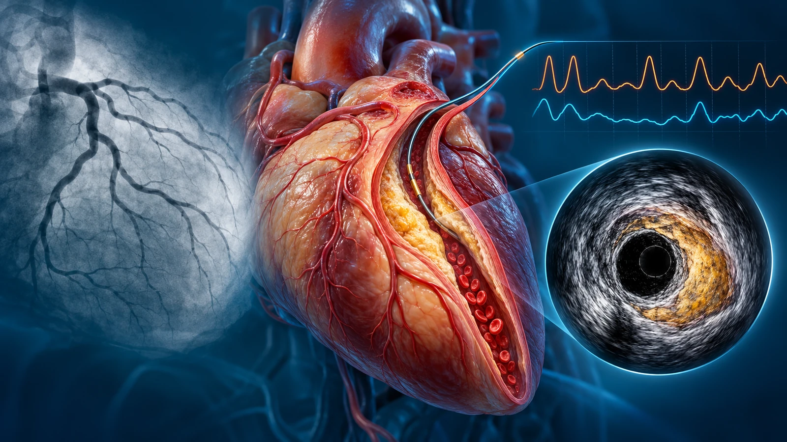
A coronary narrowing seen on angiography does not automatically mean you need a stent. In selected patients, fractional flow reserve (FFR) or iFR can help determine whether an intermediate narrowing is physiologically important, while intravascular ultrasound (IVUS) shows the artery wall, plaque and vessel dimensions and can guide stent placement. Decisions also depend on symptoms, coronary anatomy, other test results, overall risk and the likely benefit of medical therapy, PCI or bypass surgery. New or severe chest pain requires emergency evaluation rather than a routine second opinion.
Your Angiogram Shows a Blockage — Is It Actually Limiting Blood Flow? | FFR vs IVUS
Dr. Mehdi Shahriyari, Interventional Cardiologist in Dubai, explains why a coronary angiogram does not always show whether a narrowing limits blood supply, how FFR and iFR assess coronary physiology, what IVUS shows inside the artery, and why not every blockage needs a stent. Educational information only; new or severe chest pain requires emergency care.
Loading this video connects to YouTube, which may receive your IP address and browser details.
Key takeaways
- A visible coronary narrowing does not automatically require angioplasty or stenting.
- An angiogram shows the contrast-filled coronary lumen and anatomy rather than directly measuring the narrowing's functional impact.
- FFR is a pressure-derived index measured during hyperaemia, while iFR is a pressure-derived index measured at rest.
- FFR and iFR thresholds can guide selected stable coronary decisions, but results must be interpreted in clinical context.
- IVUS provides ultrasound views of the artery wall, plaque and vessel size and can help optimize selected stent procedures.
- FFR/iFR and IVUS answer different, complementary clinical questions and are not required for every patient.
- Medication, risk-factor management and follow-up remain important even when a coronary stent is implanted.
- Some complex coronary patterns may require consideration of bypass surgery rather than PCI alone.
- If symptoms suggest a heart attack or another emergency, seek urgent care; the UAE ambulance number is 998.
Section 01
A Blockage on an Angiogram Does Not Always Mean You Need a Stent
Being told that an angiogram has found a blockage in a heart artery can be unsettling. You may hear a number, 50%, 70%, perhaps more, and immediately wonder whether you need a stent. You may also wonder whether it is safe to wait, whether medicines are enough, or whether the narrowing could cause a heart attack.
These are reasonable questions. When I discuss coronary angiography results with patients, I try to begin with a more useful question than the percentage alone: What does this finding mean for your symptoms, the blood supply to your heart, and your overall health?
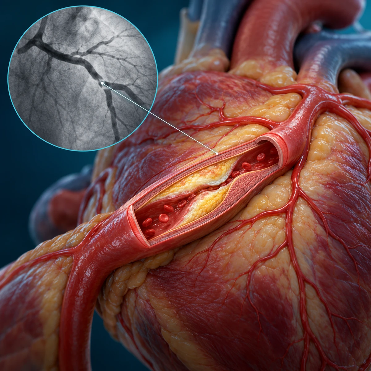
A coronary artery supplies oxygen-rich blood to part of the heart muscle. Over time, atherosclerotic plaque can build up within the artery wall and narrow the channel through which blood travels. Coronary angiography allows us to see that channel. During the procedure, a catheter is guided into an artery, usually through the wrist or sometimes the groin. Contrast dye is injected while X-ray images outline the coronary arteries.
The angiogram is extremely valuable. It shows where a narrowing lies, which vessels are involved and whether the overall anatomy suggests a potentially serious pattern of disease. However, it mainly gives us an anatomical picture of the contrast-filled lumen. It does not directly measure how much blood reaches the heart muscle during increased demand, or whether a particular narrowing explains every symptom.
Why can appearance be misleading? A narrowing can look different depending on the viewing angle, the shape of the vessel, vessel overlap and the distribution of plaque. A single visual percentage is therefore an estimate, not a complete decision. A narrowing described as “moderate” may have a meaningful physiological effect; another that appears severe may need interpretation in the context of symptoms, anatomy and other tests.
There is another distinction I want patients to understand: having coronary plaque is not identical to having a narrowing that significantly restricts blood supply. Plaque itself matters because it reflects coronary artery disease and future risk, even when a stent is not indicated. This is one reason preventive treatment remains important after an angiogram, whether or not a procedure is performed.
When assessing a stable patient, I consider whether discomfort occurs during exertion, whether it improves with rest, what previous investigations show, and whether medication has been tried or optimized. Diabetes, kidney function, heart function, the pattern of coronary disease and the patient’s priorities may all influence the decision. Sometimes the picture is clear. Sometimes we need a better understanding of what the narrowing is actually doing.
Importantly, this discussion concerns stable, planned assessment. If someone develops new, severe, persistent or worrying chest pain, especially with breathlessness, sweating, nausea or fainting—the priority is urgent emergency assessment, not arranging an elective FFR test or a second-opinion appointment. In the UAE, call 998 for an ambulance if a medical emergency is suspected.
Section 02
FFR, iFR and IVUS: Different Tools for Different Questions
Once we know what an angiogram shows, the next issue is whether additional testing could change the treatment decision. Two tools that patients increasingly encounter are fractional flow reserve (FFR) and intravascular ultrasound (IVUS). They may be used in the same catheterization laboratory, but they do not do the same job.
What does FFR measure?
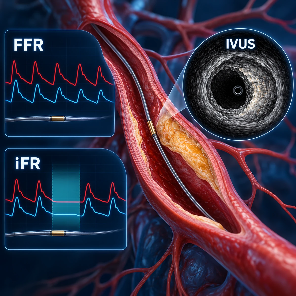
FFR is a pressure-derived measurement used to assess the physiological significance of a coronary narrowing. In selected cases, a very fine pressure-sensing guidewire is passed beyond the area of concern. Pressure on either side of the narrowing is compared while medication, commonly adenosine or another appropriate agent, produces maximal increase in coronary blood flow. This is called hyperaemia.
The resulting value helps us judge whether the narrowing is producing a meaningful pressure drop under those testing conditions. FFR does not directly measure the amount of blood reaching the heart muscle. It is an established pressure-based index that helps us understand whether the particular epicardial artery narrowing may be limiting supply.
For appropriately selected intermediate lesions in stable coronary disease, an FFR value of 0.80 or below is commonly used to indicate physiological significance. A value above 0.80 can support deferring PCI for that lesion when the measurement is reliable and the rest of the clinical picture agrees. But 0.80 is not a magic switch that automatically orders a stent. Borderline results, complex anatomy, severe left-main disease, acute presentations and other clinical factors may require a more individualized interpretation.
How is iFR different?
Instantaneous wave-free ratio (iFR) is another pressure-based index. It is measured during a particular part of the resting cardiac cycle, without the medication-induced hyperaemia used in standard FFR testing. A threshold of 0.89 or below is commonly used in clinical decision-making. As with FFR, the number must be considered alongside symptoms, coronary anatomy and other evidence; it does not make the treatment decision on its own.
For example, if an angiogram shows an intermediate narrowing, physiology can sometimes help distinguish a lesion that is likely to matter from one where stenting may add little benefit. In other situations—especially when anatomy or the clinical presentation is already decisive—these measurements may not be necessary or may be less suitable.
What does IVUS show?
IVUS answers a different question: What does the inside of the artery look like?
With IVUS, a miniature ultrasound transducer mounted on a catheter travels inside the coronary artery. Rather than showing only the outline of the contrast-filled channel, it produces cross-sectional images of the artery and its wall. These images can help assess the vessel’s dimensions, plaque burden, calcium distribution and the characteristics and length of a diseased segment.
That information becomes particularly useful when coronary angioplasty and stenting are being planned. IVUS may help the operator choose an appropriate stent diameter and length, prepare a heavily calcified lesion and assess the result after the stent has been expanded. We can look for inadequate expansion, incomplete contact with the vessel wall, or certain problems at the stent edges that might not be obvious from angiography alone.
Current evidence supports intravascular imaging guidance in selected complex coronary interventions. Optical coherence tomography (OCT) is another intracoronary imaging method that may be used for similar purposes, although it works differently from ultrasound. Neither IVUS nor OCT guarantees that a procedure will be free of complications or that future coronary events cannot occur.
So which test is better—FFR or IVUS?
I would not describe them as competing tests. FFR and iFR help answer whether a narrowing is physiologically significant; IVUS helps show what the vessel and plaque are like and can guide how a stent is implanted. In a selected patient, these pieces of information complement one another. In another patient, one may be useful and the other unnecessary. Some patients need neither.
There is also an important limit to remember: if pressure testing does not show a flow-limiting large-vessel narrowing, persistent chest discomfort still deserves attention. Some patients have symptoms related to dysfunction of the small coronary vessels, coronary spasm or causes outside the heart. A reassuring FFR result for one lesion does not mean that every possible explanation for symptoms has been excluded.
The purpose of additional testing is not to collect as many numbers or images as possible. It is to answer a clinical question that may meaningfully change what we recommend.
Section 03

From Test Results to Treatment: What Should Happen Next?
After reviewing the angiogram, and FFR, iFR or IVUS when they are appropriate, the most important decision is still personal: Which treatment strategy offers a meaningful benefit for this patient?
For many people with chronic or stable coronary disease, the foundation is guideline-directed medical therapy and long-term risk reduction. Depending on the individual, this may include cholesterol-lowering treatment, blood-pressure and diabetes management, medicines for angina, smoking cessation, suitable physical activity and attention to diet and other risk factors. The precise medicines depend on the diagnosis and clinical circumstances, so patients should not start, change or stop cardiac drugs on the basis of an online article.
When might a stent help? Percutaneous coronary intervention (PCI), also called coronary angioplasty, may be appropriate when a significant coronary narrowing is causing persistent, limiting symptoms despite suitable medical treatment, or in specific higher-risk clinical or anatomical situations. The intended benefit must be clear. For some stable patients, the principal expected benefit is improved angina and quality of life; it is not correct to promise that a stent will automatically prevent a heart attack or prolong life in every case.
The decision is different when a patient has an acute coronary syndrome, particularly a heart attack caused by an acutely obstructed artery. Urgent invasive assessment and coronary reperfusion may be required. Time-sensitive emergency care should not be delayed in order to follow the pathway designed for a stable outpatient with an intermediate narrowing.
What about bypass surgery? Some patterns of disease, [1][2][3][4][5][6]notably certain left-main or complex multivessel coronary disease, particularly in the presence of relevant additional factors—may be better considered for coronary artery bypass grafting (CABG). For complex decisions, discussion by a multidisciplinary Heart Team may help clarify the safest and most appropriate strategy. This is why it is too simplistic to ask only, “Stent or no stent?”
And what if no stent is recommended? That is not the same as saying nothing is wrong or that follow-up is unnecessary. Coronary plaque still calls for attention to risk factors. Angina can sometimes occur even without a severely obstructed large coronary artery. Persistent symptoms may lead us to consider microvascular dysfunction, coronary vasospasm or another diagnosis, depending on the history and findings.
When additional catheter-based testing is being considered, I also discuss its possible limitations and risks. Pressure-wire assessments, intravascular imaging and angiography involve invasive instruments, and the overall procedure carries risks that may include bleeding, arterial injury, rhythm disturbance, contrast-related reactions or kidney complications in susceptible patients. Serious complications are uncommon in appropriately selected settings, but they cannot be described as impossible. Whether any additional test is worth doing depends on whether its result is likely to improve the decision.
Five questions to ask when reviewing your angiogram
- Which artery is narrowed, and how certain are we about its significance?
- Could FFR, iFR or IVUS add information that changes my treatment?
- What result or symptom is the proposed treatment intended to improve?
- What are the reasonable alternatives, medication, PCI or, in selected cases, bypass surgery?
- What treatment and follow-up will I still need after this decision?
If you are in Dubai and have a previous coronary angiogram, the report is useful, but the actual images and your clinical history can be equally important for a considered second opinion. For stable, non-emergency concerns, a review can help clarify the recommendation and its alternatives. It should never become a reason to postpone treatment for a suspected heart attack.
My message is simple: an angiogram gives us an important picture; the best decision comes from understanding what that picture means for you.
Dr. Mehdi Shahriyari
Interventional Cardiologist, Dubai, UAE
Medical information is for patient education and does not replace personalized assessment. If you have new, severe or concerning chest pain now, seek emergency care; in the UAE, call 998.
Patient questions
Patient questions
Q1. Does a 70% blockage on an angiogram automatically mean I need a stent?
No. The percentage is only one part of assessment. Symptoms, coronary location, overall anatomy, clinical urgency, medication response and, when appropriate, physiological testing help determine whether PCI offers a useful benefit. Some higher-risk patterns need a different strategy.
Q2. What is the difference between FFR and IVUS?
FFR uses a pressure-sensing guidewire during maximal hyperaemia to assess the physiological significance of a narrowing. IVUS uses an ultrasound catheter to examine the vessel wall, plaque and dimensions. They answer different questions and may be complementary.
Q3. What FFR result suggests a narrowing is significant?
For selected intermediate coronary lesions in stable disease, FFR at or below 0.80 is commonly used to identify physiological significance. An FFR above 0.80 may support deferring PCI in the appropriate context. Borderline values and individual clinical circumstances require careful interpretation; the number alone does not prescribe a stent.
Q4. Is iFR the same as FFR?
They are related pressure-based physiological assessments, but iFR is measured at rest without the medication-induced hyperaemia used for conventional FFR. A value at or below 0.89 is a commonly used iFR threshold. Your clinician selects and interprets the test according to the clinical situation.
Q5. Can IVUS make a coronary stent safer or more accurate?
IVUS can help assess vessel dimensions, plaque and whether a stent is adequately expanded and apposed. Evidence supports intravascular imaging guidance in selected complex PCI procedures, but it is not necessary in every case and cannot guarantee the absence of complications.
Q6. If FFR is not abnormal, why might I still have chest pain?
A non-flow-limiting result for one epicardial narrowing does not rule out all cardiac or non-cardiac causes of chest pain. Coronary microvascular dysfunction, vasospasm or other conditions may need consideration. New, severe or concerning chest pain warrants urgent emergency evaluation rather than waiting for outpatient investigation.
Dubai / UAE context
Dubai / UAE context
In Dubai, people who have undergone coronary angiography may seek clarification about a reported narrowing, whether physiological testing such as FFR or iFR would add value, and whether IVUS-guided PCI is appropriate. For stable, non-emergency decisions, a cardiology assessment or second opinion should review symptoms, current medicines, available angiographic images and reports, prior investigations and the patient's preferences. Access and availability of specific investigations vary by facility and indication; not every patient needs an additional invasive test. If symptoms suggest a heart attack or another medical emergency, seek emergency assessment immediately rather than arranging a clinic visit. In the UAE, call 998 for an ambulance.
Evidence & sources
Evidence & sources
Related reading
Related reading
 ▶ Video
Chest Pain: Heart or Acid Reflux?
Chest pain can come from the heart, acid reflux or several other causes. Learn the clues doctors consider, the tests that may be needed, and when chest pain requires urgent care.

Chest Pain in Dubai: Heart Attack Symptoms, Warning Signs, and When It Is an Emergency
Learn the warning signs of heart attack, when chest pain becomes an emergency in Dubai, and when recurring symptoms should be assessed by a cardiologist.
Discussion & comments
Discussion & comments
Comments are moderated. Do not post urgent medical questions, personal identifiers or private medical records.